
Study

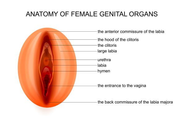
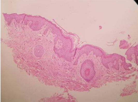
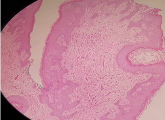
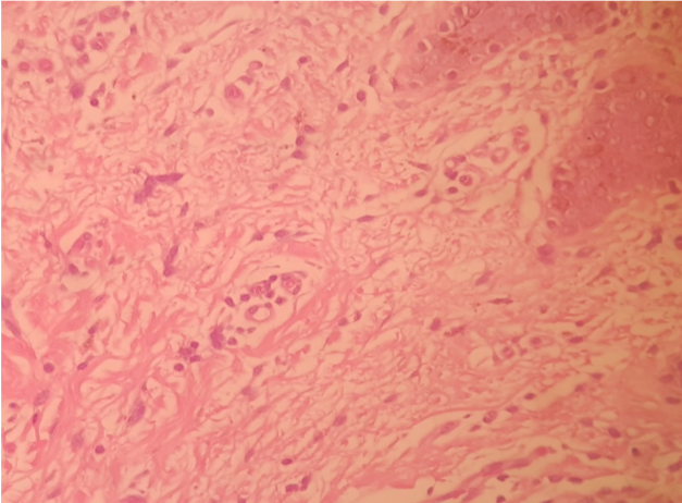
Histology of Biopsies of Labia Majora taken by punch on Patients treated by Endopeel

Alain Tenenbaum

Endopeel Inventor

Mauro Tiziani

Endopeel Co-Inventor

Juan Barcia

Anatomo Pathologist
anatomy of female genital organs.jpg
30 minutes post endopeel +PRP on large labia

30 minutes biopsy post endopeel on Labia Majora

30 and 60 minutes after the injection of Endopeel :

  • no notable changes were observed.

  • Absence of necrosis in all layers of the skin of the large labia

  • no thrombosis

  • no hemorrhage  were observed

30 days biopsy post endopeel +PRP on large labia

30 days biopsy post endopeel on Labia Majora

30 days after treatment

  • a greater cellularity was observed in the dermis (fibroblasts and mononuclear cells)

  • in conjunction with a mild collagen fibrosis in different planes (disordered)

60 days biopsy post endopeel +PRP on large labia

60 days biopsy post endopeel on Labia Majora

60 days after treatment

  • the angiogenic process is at its highest level

  • with high density of small neoformed capillaries in the middle and upper dermis.

  • Fibrosis has become ordered by remodeling phenomena.

  • No tissue damage (necrosis, hemorrhage, etc.)

MACRO :60 days biopsy post endopeel +PRP on large labia

MACRO :60 days biopsy post endopeel on Labia Majora